Ankle mobility, especially dorsiflexion (bringing your toes up towards your head), is very important at all scales of movement practice. At one end, restricted dorsiflexion means that you’ll have less time with your whole foot on the floor as you walk, making you less stable and more prone to falling. At the other end, when working on deep squats and advanced squat variations, or any kind of low-to-the-ground movement, dorsiflexion range is almost always the main restriction you’ll face (along with restrictions in hip flexion).
I already wrote a blog post earlier this year where I present a few stretches for this issue, and today we’ll explore some anatomy and a few images to promote a good dorsiflexion range.
A bit of anatomy
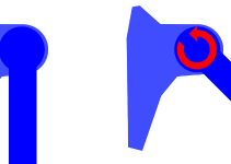
The ankle joint, linking the tibia and fibula to the foot (the talus being the bone at the top of the foot, articulating with the leg), is a sort of wheel (talus) rolling inside a fork formed by the tibia and fibula (or when your foot is on the ground, the fork rolling on top of the wheel). See this post for another description of the same area. Another detail that we will consider today is that the wheel is wider at the front, so that the fork also has to get wider when it goes forward (dorsiflexion). See the video below around 1:45 to get an idea of the movements involved. The fork is held together by a membrane joining the tibia and fibula (interosseous membrane), and a few ligaments joining the ends of the tibia and fibula to the foot (around 1:50).
The last piece of the puzzle that we will consider is the tendons of muscles located deep in the shin and acting in the foot, that pass along the heel bone (calcaneus), kept in place by grooves in the bone.
One source of restriction to dorsiflexion is tightness in the calf muscles, and this is addressed in the aforementioned post. The other sources are tightness in the interosseous membrane and ligaments I mentioned above (this can be helped by massage and other manual work), and less-than-optimal movement habits. The imagery I present here helps with both these factors, restoring better movement patterns that will, in turn, help release tightness in the affected tissue.
Images
The first image is simply to think of your heel going back and slightly down as you’re flexing your leg (think plié). You can complement this image with your calf muscles elongating (using an image similar to that presented at the end of this post) and the plantar fascia (the horizontal springs in this post) stretching.
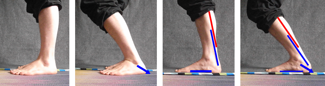
Another layer you can add is to see the slings formed by the tendons of the deep muscles mentioned above loosening to give more space for the heel to go back and away from the ankle.

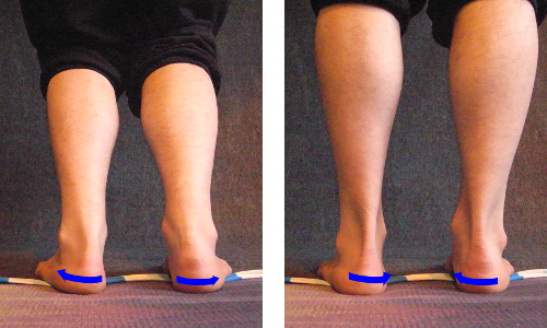
The next image involves movements inside the foot, about which I will not goin into anatomical detail. It consists in seeing your heel turn out when you flex, and turn in again when you come back to neutral. You wil probably feel that this movement affects your arch, and that’s perfectly normal. You can even use this image to correct your arch if it’s not ideal: if your arch is a bit flat, emphasise the “turning back in” part when you come back to neutral, and if your arch is a bit deep, emphasise the “turning out” part when you flex.
I hope this helps you in all your movement endeavors!
Continue reading: Ankle Mobility, Foot Posture And Alignment, A Key To Flexibility Training.
Want more like this?
Check out the following blogs from massage therapists I know from around London:- On The Run Health and Fitness on running, nutrition and sports massage.
- The Soma Room on sports massage and exercise.

No Responses