The foot is our literal basis when we stand (or squat), yet most people know very little about it. This week we start solving this issue! I will guide you through a small exploration of your foot, to get to know some of its structures, and to get some awareness of its possibilities. Some of it might be a bit sore, which simply indicates that these areas could get more movement (as in, move more often and in more different ways).
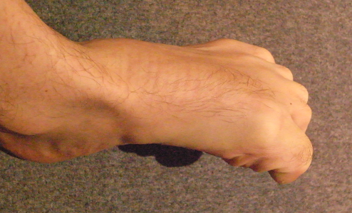
Let’s start with an easy one: the calcaneus, or heel bone. Spend a few minutes exploring its shape, size and position. Start close to the heel, find the very hard surface under the skin, and trace it up and forward. On your way up you’ll meet the Achilles tendon; try and find the top of the calcaneus between your Achilles and your tibia (shin-bone). Going forward on the sides of the foot, find where it ends; you’ll feel a serious dip into softer tissue.
Now for a slightly deeper, but no less important bone: the talus. This is the point of contact between the foot and the leg. It’s also the “keystone” to the medial longitudinal arch of the foot (“the” arch). Grasp your foot on either side, just in front of the calcaneus, and walk your way up. You’ll find a rigid body, and that’s your talus. Your fingers should be just under your malleoli, and that’s no coincidence: the malleoli are the end of the tibia and fibula (lower leg bones), grasping the talus and gliding on top of it.

Now let’s start at the other end. I’m sure you don’t need any pointers to identify your toes, but spend some time exploring them. They’re exactly like your fingers: three phalanges (segments) for the 4 smaller ones, and two for the big one. Locate the nine interphalangeal (between the phalanges) joints of the toes of each foot. And now, try and locate the joints between the toes and the rest of the foot. Hint: they’re way further in than you think!
From the toes, let’s find the metatarsals; those are the “roots” of the toes: bones that run more or less parallel to the toes, towards (or from, depending on perspective) the ankle. Try and find those bones, and more importantly, the spaces between them (that might be a bit sore). In particular, pay attention to the shape of the fifth metatarsal, the one supporting the pinky toe. Its proximal (towards the body) end is very distinctive.
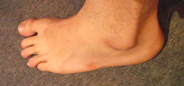
I sit cross-legged a lot, hence the area of thicker skin exactly at the end of the fifith metatarsal.
Our last bony landmark will be the navicular, the easiest to locate among the 5 remaining bones (we already touched on 21!). First locate the dip between your leg and you foot, on the front of your ankle, slightly towards your midline. From there, obviously if you go towards the leg, you’ll find the tibia; and if you go in the other direction, you’ll find our friend navicular.

Now a few soft tissue points of interest.
We already mentioned the Achilles tendon at the back, down from the powerful calf muscles, and attaching to the calcaneus. Now we’ll go meet it’s (almost) counterpart on the front: the tendon of the tibialis anterior, the main muscle opposing plantar flexion (preventing your foot from slamming into the floor when you walk or run, especially downhill). For this, just dorsiflex your foot (take the end of your foot towards your head) while relaxing your toes. It will be the only prominent tendon of the front of the ankle. If you can’t relax your toes in this position, look for the tendon closest to your midline.
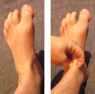
The tendons of the extensors of the toes are easy to locate (just lift your toes). Try to also find them with relaxed toes; notice how they feel when relaxed, and when under tension. In either state, follow them as far up (towards your ankle) as you can.
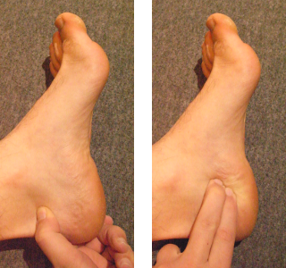
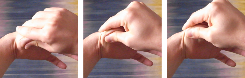
Lastly, let’s try and locate the medial border of your plantar fascia (possibly two different layers, depending on the state of your tissue): put your fingers on the inside of the foot, just close enough to the sole that you’re not just over a bone. Dorsiflex your foot and lift your toes. Then gently travel towards your sole with your fingers. You’ll meet a hard line, kind of like a tendon. This is the medial border of your plantar fascia! If you do a lot with your feet, you might notice a rather thick one, very close to the malleoli, and a thinner one, closer to the sole. Those are two layer of the plantar fascia, playing slightly different roles in your stability, strength and “jumpyness”.

Here you can actually see the two layers of plantar fascia.
I hope you learned something this week, and that your feet will be happy from the attention. Please share this post with your friends, and subscribe to the newsletter to know when a new post goes online.
Continue reading: Self-Massage For The Feet, Explore Your Knee, Foot Posture And Alignment.
Want more like this?
Check out the following blogs from massage therapists I know from around London:- On The Run Health and Fitness on running, nutrition and sports massage.
- The Soma Room on sports massage and exercise.


No Responses